Articles

Dr. Holland Optometrists Blog

Dr. Holland Optometrists Blog is your trusted source for informative articles, expert insights, and valuable tips related to eye health and vision care. Explore a wealth of knowledge on topics ranging from eye conditions, vision correction options, to maintaining optimal eye wellness. Stay informed and prioritize your eye health with our engaging and educational blog content.

Optical Coherence Tomography

Optical Coherence Tomography

Optical Coherence Tomography is a non-invasive imaging test that may be performed as a standard part of your regular, comprehensive exams, or you may be able to request this test as an addition to your usual exam.

Optical Coherence Tomography uses light waves to take cross-section images of your retina, which is the area of light-sensitive cells at the back of your eye that is responsible for receiving light and transmitting it into messages that are sent up to the brain. The technology behind OCT enables your eye doctor to see each of the different layers that make up the retina. By being able to see these and measure them, they can obtain a much clearer picture of the overall health and condition of your eyes.

Why are Optical Coherence Tomography scans important?

When you choose to have an OCT scan at fairly regular intervals, such as during your normal comprehensive eye exams, your eye doctor can compare newer results to previous ones. This helps them to build up a picture of the health of your eyes, and spot any changes which may be concerning, early, before they cause symptoms or have a permanent effect on your vision. 

Anyone can have an OCT scan, but they are particularly recommended for patients over the age of 25 who are concerned about the health of their eyes, or who are at risk of or already have diabetes, glaucoma or a family history of eye disease. This is because they can be used to spot the early signs of a range of eye diseases, including glaucoma, diabetic retinopathy, macular degeneration, disorders of the optic nerve and more – even before you realize that you are affected.

Read more
Senior Care

Senior Care

Senior eye care is crucial for maintaining optimal vision and overall eye health as we age. As individuals grow older, they become more susceptible to age-related eye conditions. Regular comprehensive eye exams play a key role in early detection and management of these conditions, helping to preserve vision and prevent potential vision loss. By staying proactive with eye care and seeking timely professional assistance, seniors can enjoy clear, comfortable vision and ongoing eye health for years to come.

Common Eye Conditions in Seniors

As we reach our golden years, there are several eye conditions that become more prevalent. These conditions can have a significant impact on our vision and overall quality of life.

  • Presbyopia: This condition affects near vision, making it difficult to focus on close objects. It is a normal part of aging and is typically addressed with reading glasses or multifocal lenses.

  • Floaters and Flashes: As the vitreous inside the eye changes with age, it can lead to the perception of floaters (small dark spots or lines) and flashes of light. While often harmless, sudden onset of floaters and flashes may indicate a retinal tear or detachment and should be promptly evaluated.

  • Age-Related Macular Degeneration (AMD): AMD is a leading cause of vision loss in older adults. It affects the macula, the central part of the retina, leading to blurred or distorted central vision. There are two forms of AMD, dry AMD, and wet AMD, with the latter being more severe.

  • Cataracts: Cataracts are a common age-related condition characterized by the clouding of the eye's natural lens. This can lead to blurry vision, glare, and difficulty seeing in low-light conditions.

  • Glaucoma: Glaucoma is a group of eye conditions that damage the optic nerve, often due to elevated intraocular pressure. It can result in peripheral vision loss and, if left untreated, may lead to total blindness.

  • Diabetic Retinopathy: For seniors with diabetes, diabetic retinopathy is a concern. It occurs when high blood sugar levels damage the blood vessels in the retina, leading to vision problems and potential blindness if not managed.

  • Dry Eye Syndrome: Seniors may experience dry eye syndrome, characterized by insufficient lubrication and moisture on the surface of the eye. It can cause discomfort, redness, and fluctuating vision.

Read more
Stellest Lenses

Stellest Lenses

In September 2025, the FDA approved Stellest lenses for children ages 6 to 12 to correct and manage myopia, a major milestone for myopia care. These lenses have been successfully used in countries across Europe, Canada, and Asia since 2020, showing promising results in slowing eye growth associated with myopia.
 

The Importance of Early Myopia Management

Myopia typically begins in early childhood, often between the ages of 6 and 10, and tends to worsen quickly during the school years as children spend more time reading and using digital devices. The sooner it begins, the higher the risk that it will progress to more severe levels. Every increase in myopia level significantly raises the risk of eye health complications later in life, such as retinal detachment or glaucoma.

By beginning myopia management as soon as possible, parents can help slow eye growth and protect long-term vision. With innovative options like Stellest lenses, families now have effective, science-backed solutions to take control of their child’s myopia.
 

What Are Stellest Lenses?

Stellest lenses are a groundbreaking innovation in myopia control, developed by Essilor. They look like regular eyeglass lenses but are designed with specialized optical technology to both correct vision and slow the progression of myopia.

Read more
Vision Therapy: Conditions

Vision Therapy: Conditions

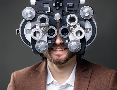
Vision therapy is a specialized form of treatment that aims to improve visual function and performance. It involves a series of eye exercises, activities, and visual training techniques designed to address a wide range of vision-related issues. Unlike traditional eye care, which often focuses on correcting refractive errors with glasses or contact lenses, vision therapy takes a holistic approach to addressing the underlying causes of visual problems.

By targeting the root causes of these issues, vision therapy can help individuals achieve better visual function, improve their overall well-being, and enhance their ability to perform daily tasks with greater ease and efficiency.


Amblyopia


Amblyopia, commonly known as "lazy eye," is a common vision condition that affects approximately 2-3% of the population. It occurs when one eye develops reduced visual acuity due to the brain's inability to properly process information from that eye.

In many cases, amblyopia develops during childhood, often due to factors such as refractive errors, strabismus (misaligned eyes), or visual deprivation. If left untreated, amblyopia can lead to permanent vision loss in the affected eye, as the brain increasingly favors the stronger eye and the weaker eye's visual processing abilities deteriorate.

Vision therapy is a highly effective treatment for amblyopia, as it aims to retrain the brain to properly utilize the weaker eye. Through a series of specialized exercises and activities, vision therapy can strengthen the neural pathways between the brain and the affected eye, improving visual acuity and binocular vision.

The key to successful treatment lies in the brain's remarkable ability to adapt and change, known as neuroplasticity. By engaging in targeted vision therapy, individuals with amblyopia can stimulate this neuroplasticity, allowing the brain to reorganize and improve its processing of visual information from the weaker eye.


Strabismus


Strabismus, also known as crossed eyes or misaligned eyes, is a condition where the eyes do not properly align with each other. This can cause one eye to turn inward, outward, upward, or downward, resulting in a lack of binocular vision and depth perception.

Strabismus can have a significant impact on an individual's visual function, self-esteem, and overall quality of life. Left untreated, it can lead to the development of amblyopia (lazy eye) and other vision-related problems.

Vision therapy is a highly effective treatment for strabismus, as it focuses on retraining the brain and the eye muscles to work together in a coordinated manner. Through a series of specialized exercises and activities, vision therapy can help individuals with strabismus improve their eye alignment, binocular vision, and depth perception.


Binocular Vision Disorders


Binocular vision disorders are a group of conditions that affect the way the eyes work together as a team. These disorders can result in a range of visual problems, including difficulty with depth perception, eye strain, headaches, and difficulties with reading and other visual tasks.

Some common binocular vision disorders that can be effectively treated with vision therapy include:

  • Convergence Insufficiency: This condition is characterized by the inability of the eyes to properly converge, or come together, when focusing on a close object. This can lead to eye strain, headaches, and difficulties with reading and other near-work activities.

  • Accommodative Dysfunction: Accommodative dysfunction is a problem with the eye's ability to focus, or "accommodate," on near objects. This can cause blurred vision, eye strain, and difficulties with reading and other close-up tasks.

  • Vergence Dysfunction: Vergence dysfunction refers to a problem with the eye's ability to work together to maintain proper alignment and focus. This can lead to double vision, eye strain, and difficulties with depth perception.

Through vision therapy, individuals with binocular vision disorders can retrain their visual system to work more efficiently and effectively. The goal of vision therapy is to improve the coordination and synchronization of the eyes, ultimately leading to better visual performance and reduced visual symptoms.

Read more
Presbyopia

Presbyopia

Presbyopia is a natural and inevitable part of aging that affects the eyes' ability to focus on close objects. Unlike other vision conditions, presbyopia isn’t caused by the shape of the eye or structural abnormalities but is instead a result of the eye’s lens losing its flexibility over time.

What is Presbyopia?

Presbyopia is a refractive error, meaning it affects how the eyes bend (or refract) light to focus it on the retina. Unlike nearsightedness (myopia) or farsightedness (hyperopia), which are caused by the shape of the eyeball or cornea, presbyopia is due to the aging of the eye's lens. Over time, the lens becomes less flexible, making it difficult to focus on nearby objects. Presbyopia isn’t a disease or an abnormality—it’s simply a natural consequence of the aging process.

What Causes Presbyopia?

The lens inside the eye changes shape to focus light on the retina, allowing us to see clearly at varying distances. When viewing nearby objects, the lens thickens and curves to increase its refractive power.

As we age, however:

  • Lens Stiffening: The lens loses elasticity, making it harder to bend and adjust for near vision.

  • Weakened Ciliary Muscles: The muscles that help change the shape of the lens may lose their strength over time, further reducing the eye’s ability to focus on close objects.

  • Structural Changes: The lens also grows thicker and less transparent with age, which contributes to reduced focusing ability.

These changes are gradual, typically starting in a person’s 30s and becoming noticeable by their early to mid-40s.

Read more
Surgical Co-Management

Surgical Co-Management

In the realm of eye care, surgical co-management has emerged as a collaborative approach that aims to provide patients with comprehensive and seamless treatment. This concept involves the joint efforts of optometrists and ophthalmologists, each bringing their unique expertise to the table. By working together, these eye care professionals strive to enhance patient outcomes and deliver exceptional care.

The Role of Optometrist and Ophthalmologist in Surgical Co-Management

Surgical co-management is built upon the unique skill sets and areas of expertise of optometrists and ophthalmologists. By understanding their respective roles, you can appreciate the synergy that this collaborative approach fosters.


Optometrists are primary eye care professionals who specialize in the examination, diagnosis, and non-surgical treatment of vision disorders. Their responsibilities in surgical co-management include:

  • Performing comprehensive eye examinations and evaluations

  • Monitoring and managing pre-existing eye conditions

  • Providing pre-operative and post-operative care

  • Educating patients on surgical procedures and aftercare

  • Collaborating with ophthalmologists to ensure continuity of care

Ophthalmologists are medical doctors who specialize in the diagnosis, treatment, and surgical management of eye diseases and disorders. Their role in surgical co-management encompasses:

  • Evaluating patients' candidacy for surgical interventions

  • Performing complex surgical procedures

  • Providing specialized medical and surgical care

  • Collaborating with optometrists to ensure seamless patient care

  • Monitoring and managing post-operative complications

  • By combining the expertise of optometrists and ophthalmologists, surgical co-management ensures that patients receive comprehensive and coordinated care throughout their treatment journey.

How Surgical Co-Management Works

Surgical co-management is a well-orchestrated process that involves several key steps. Understanding how it works can help you navigate this collaborative approach with confidence.

  • Initial Evaluation: The process typically begins with an optometrist conducting a comprehensive eye examination. During this evaluation, the optometrist assesses the patient's visual needs, identifies any potential issues, and determines if a surgical intervention is necessary.

  • Referral and Consultation: If surgery is recommended, the optometrist refers the patient to an ophthalmologist for further evaluation and consultation. This step ensures that the patient receives specialized medical advice and a thorough assessment of their suitability for the proposed surgical procedure.

  • Pre-operative Care: The optometrist plays a crucial role in providing pre-operative care, which may include managing any existing eye conditions, ensuring the patient understands the surgical process, and addressing any concerns or questions they may have.

  • Surgical Procedure: The ophthalmologist performs the necessary surgical intervention, leveraging their specialized training and expertise in surgical techniques.

  • Post-operative Care: After the surgery, the patient's care transitions back to the optometrist, who closely monitors the recovery process and provides post-operative care and management. This may involve follow-up appointments, monitoring for any complications, and ensuring the patient adheres to the prescribed treatment plan.

  • Ongoing Collaboration: Throughout the entire process, the optometrist and ophthalmologist maintain open communication and collaborate closely. This ensures that the patient's care is seamless, and any concerns or issues are promptly addressed by the appropriate healthcare professional.

Read more
Optomap

Optomap

Optomap is an innovative new technology that gives eye doctors the ability to perform ultra-wide retinal imaging that is far superior to what can currently be achieved using conventional retinal imaging options. In contrast to conventional retinal imaging, Optomap captures at least 50% more of the retina in a single capture, and with Optomap’s multi-capture function, up to 97% of the retina can be viewed. This gives eye care professionals greater opportunity to monitor the health and condition of patient vision.

Why is Optomap important?

Optomap is another great preventative eyecare technology tool. By allowing your eye doctor to have a comprehensive view of your retina, they will be able to detect any developing eye diseases early on, before they have a detrimental impact on your vision and day to day life. Not only can Optomap detect eye conditions such as retinal holes, retinal detachment, macular degeneration and diabetic retinopathy, but it can also be used to identify some general health conditions such as cardiovascular disease, stroke and cancer.

Read more
Medical Eye Exam

Medical Eye Exam

Medical eye exams are comprehensive evaluations of the health and function of the eyes, essential for maintaining optimal vision and preventing potential eye conditions. Regular medical eye exams are fundamental for proactive eye health management and ensuring ongoing visual wellness.

What is a Medical Eye Exam?

A medical eye exam is a comprehensive examination of your eyes conducted by an optometrist. This examination goes beyond a routine vision test and delves into the overall health of your eyes. During a medical eye exam, various tests will be performed to assess your visual acuity, peripheral vision, eye movement, and the health of your eye structures.

Medical eye exams can detect early signs of eye conditions that may not present noticeable symptoms in their initial stages. By diagnosing these conditions early, you have a greater chance of successful treatment and preventing further deterioration of your vision.

Medical eye exams can also identify underlying health conditions that may manifest in your eyes, providing an opportunity for early intervention and management.

Common Conditions Detected During a Medical Eye Exam

During a medical eye exam, several common eye conditions can be detected, even before noticeable symptoms occur. One such condition is age-related macular degeneration (AMD), which affects the central part of the retina responsible for sharp, central vision. By examining the retina, your optometrist can identify early signs of AMD and recommend appropriate treatment to slow its progression.

Another condition that can be detected during a medical eye exam is cataracts. Cataracts occur when the lens of your eye becomes cloudy, leading to blurred vision. Through a comprehensive examination, your eye doctor can assess the severity of your cataracts and recommend the most suitable treatment option, which may include surgery to remove the cloudy lens.

Hypertension is a common health condition that can have serious consequences if left untreated. The health of your eyes can provide valuable insights into your cardiovascular health. During a medical eye exam, an optometrist can observe changes in the blood vessels of your retina, such as narrowing or leaking. These changes can indicate underlying hypertension.

Read more
Multifocal Contact Lenses

Multifocal Contact Lenses

In the ever-evolving world of vision correction, multifocal contact lenses have emerged as a game-changer for individuals seeking a seamless solution to address their presbyopia. As you navigate through life's various stages, your eyes may undergo changes that can impact your ability to focus clearly on objects at different distances. Multifocal contact lenses offer a convenient and effective way to combat this challenge, providing you with a versatile and comfortable option for correcting your vision.

What Are Multifocal Contact Lenses?

Multifocal contact lenses are specialized lenses designed to correct presbyopia, a condition that typically occurs as you age, making it difficult to focus on near objects. Unlike traditional single-vision lenses that correct either near or distance vision, multifocal contact lenses incorporate multiple prescriptions into a single lens. This innovative design allows you to enjoy clear vision at various distances, eliminating the need for separate reading glasses or bifocals.
 

How Do Multifocal Contact Lenses Work?

Multifocal contact lenses employ advanced optical technologies to provide seamless vision correction. These lenses feature distinct zones or concentric rings that correspond to different prescriptions for near, intermediate, and distance vision. As your eyes move and focus on objects at varying distances, the appropriate zone of the lens comes into play, ensuring a clear and comfortable visual experience.

There are two main types of multifocal contact lens designs:

  • Simultaneous Vision Design: In this design, the lens incorporates multiple prescriptions simultaneously, allowing your eyes to utilize the appropriate zone based on the viewing distance. This design can take some time to adjust to, as your brain learns to interpret the different visual inputs.

  • Alternating Vision Design: Also known as "segmented" or "translating" lenses, these lenses have distinct zones for near and distance vision. As you move your eyes or shift your gaze, the appropriate zone is positioned in front of your pupil, providing the necessary correction.


Eye Conditions That Multifocal Contact Lenses Can Treat

Multifocal contact lenses are primarily designed to address the following vision-related conditions:

  • Presbyopia: As you age, the natural lenses in your eyes lose flexibility, making it challenging to focus on nearby objects. Multifocal contact lenses can correct this age-related farsightedness, allowing you to see clearly at all distances.

  • Myopia (Nearsightedness): If you struggle to see distant objects clearly, multifocal contact lenses can incorporate a prescription to correct myopia, while also providing near and intermediate vision correction.

  • Hyperopia (Farsightedness): For individuals with farsightedness, multifocal contact lenses can offer a solution by incorporating a prescription to improve distance vision, along with near and intermediate vision correction.

  • Astigmatism: Some multifocal contact lenses are designed to correct astigmatism, a condition caused by an irregularly shaped cornea or lens, resulting in blurred or distorted vision.

Read more
Optikam

Optikam

Eye care professionals use Optikam’s technology to capture more than 3 million eyewear measurements every year. The OptikamPad iPad app is a total dispensing solution that enables eye care professionals to successfully assist patients at all stages of the eyewear dispensing process, providing them with a unique and custom patient experience.

Optikam Posture Devise (OPD)

You may be surprised to learn that wearing glasses can and likely will affect your posture. Glasses lenses are most accurate when you look directly through their center. This means if your glasses are sitting too low or have slipped down your nose, you may find that you are subconsciously tilting your head back and this can affect your overall posture.

Optikam’s OPD measurement device is a cutting-edge tool that obtains eyewear measurements that take into account how the frame will be worn by patients, enabling the fit to be customized to their individual parameters. The ten measurements taken into account when determining each patient’s position of wear include:

  • Monocular pupillary distance

  • Multifocal seg heights

  • Pantoscopic tilt

  • Rear vertex distance

  • Wrap (face form tilt)

  • Near pupillary distance

This results in frames that not only look fantastic, but that also fit perfectly, remaining both comfortable and stable on the face without you needing to adopt an unnatural posture. The measurements obtained by the Optikam OPD measurement tool are immediately visible on your eye doctor’s tablet so that they can recommend which alterations to the frames are needed to ensure that the frames fit with precision and gives you the best visual experience.

Read more
all eye care services
All Eye Care Services

Find personalized eye care excellence with our comprehensive services. From eye exams to advanced diagnostics, trust us for all your vision needs.

contact us
Keep In Touch

For non-urgent questions or to learn more about our services, contact us today!